Now it will be possible to assess muscle function using a new system developed at Friedrich-Alexander-Universität Erlangen-Nürnberg. Biologists have developed a system called MechaMorph that can precisely quantify muscle weakness caused by structural changes in muscle tissue.
Even more, the system does not require additional sophisticated biomechanical recordings. And it could make taking tissue samples for diagnosing myopathy superfluous.
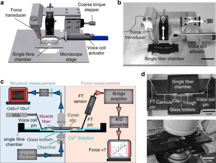
Prof. Dr. Oliver Friedrich said, “We engineered a miniaturized biomechatronics system and integrated it into a multiphoton microscope, allowing us to directly assess the strength and elasticity of individual muscle fibers at the same time as recording structural anomalies.”
With the end goal to demonstrate the muscle’s capacity to contract, the scientists dunked the muscle cells into arrangements with expanding centralizations of free calcium particles. Calcium is additionally in charge of activating muscle withdrawals in people and animals. The viscoelasticity of the filaments was additionally estimated, by extending them little by little. An exceedingly delicate indicator recorded mechanical opposition practiced by the muscle strands braced on the gadget.
Oliver Friedrich said, “The technology, however, merely the first step towards being able to diagnose muscle disorders much more easily in future: Being able to measure isometric strength and passive viscoelasticity at the same time as visually showing the morphometry of muscle cells has enabled us, for the first time, to obtain direct structure-function data pairs.”
“This allows us to establish significant linear correlations between the structure and function of muscles at the single fiber level.”
The data pool will be utilized in future to dependably anticipate powers and biomechanical exhibitions in skeletal muscle solely utilizing optical assessment dependent on SHG pictures (the initials remain for Second Harmonic Generation and allude to pictures made utilizing lasers at second consonant recurrence), without the requirement for complex strength estimations.
At present, muscle cells still must be expelled from the body before they can be analyzed utilizing a multiphoton magnifying instrument. In any case, it is conceivable this may wind up unnecessary in future if the vital innovation can keep on being scaled down, making it feasible for muscle capacity to be inspected, for instance, utilizing a miniaturized scale endoscope.
The results have been published in the renowned journal Light: Science & Application.
