Organ formation is an inherently biophysical process, requiring large-scale tissue deformations. Yet, understanding how complex organ shape emerges during development remains a significant challenge.
Fish comes in various colors, shapes, and patterns. Despite such diversity, a general feature that can be commonly found in fish such as salmon or tuna once they are served is the ‘V’ patterns in their meat. While this appears to be genetically observed in the muscle arrangement of most fish species, how such a generic ‘V’ pattern arises is puzzling.
Now scientists from NUS Mechanobiology Institute (MBI) have figured out the science behind the formation of the ‘V’ patterns in the myotome of zebrafish embryos. These ‘V’ patterns are also called chevron patterns that commonly found in the swimming muscles of fish.
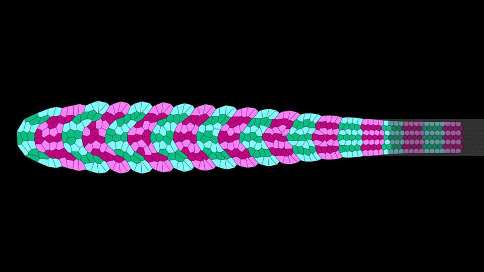
Fish’s side to side swimming motion is powered by myotome– a group of muscles served by a spinal nerve root. Initially, each future developing myotome segment is cuboidal in shape. However, over five hours, it deforms into a pointed ‘V’ shape.

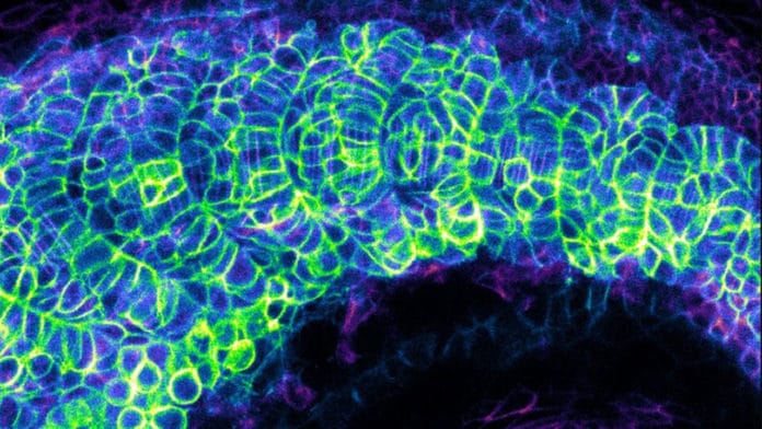
To discover how this deformation takes place, the group embraced a combination of various systems — imaging of the developing zebrafish myotome at single-cell resolution, quantitative analysis of the imaging data, and fitting the quantitative data into biophysical models.
Scientists then identified certain physical mechanisms that they thought might be guiding chevron formation during fish development.
Scientists observed that the developing myotomes are physically associated with other embryonic tissues, for example, the neural tube, notochord, skin, and ventral tissues. The strength of their connection with these various tissues changes at different time points of myotome formation, and in like manner, multiple measures of friction are created over the tissue. Adequately, the side areas of the developing myotome are under more prominent grating than the central region. As new sections push the myotome forward, this prompts the development of a shallow ‘U’ shape in the myotome tissue.
Later on, cells within the future myotome begin to elongate as they form muscle fibers. Scientists revealed that this transformation process generates an active, non-uniform force along with specific directions within the somite tissue, which results in the ‘U’ shape sharpening into the characteristic ‘V’-shaped chevron. Lastly, orientated cell rearrangements within the future myotome help to stabilize the newly acquired chevron shape.
Asst Prof Saunders, a theoretical physicist who applies physical principles to characterize biological processes that take place during development, said, “This work reveals how a carefully balanced interplay between cell morphology and mechanical interactions can drive the emergence of complex shapes during development. We are excited to see if the principles we have revealed are also acting in the shaping of other organs.”
Through this study, the MBI scientists show how temporally and spatially varying biophysical forces play a role in determining the form of an organism.
The findings of the study were published in the journal Proceedings of the National Academy of Sciences of the United States of America on 26 November 2019.
