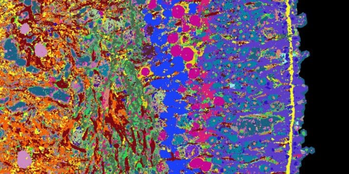
The human retina is a complex and vital part of the eye that plays a crucial role in our ability to see. Researchers from Basel and Zurich are currently working on creating a high-resolution atlas for the development of the human retina. To achieve this, they are using a new method to visualize over 50 proteins simultaneously. This breakthrough imaging technique allows them to understand the development and function of the human retina.
Researchers are creating a specialized atlas to identify cell types, genes, and proteins found in human tissue and organs, including organoids. The atlas aims to understand how tissues form during embryonic development and identify the causes of diseases. This atlas aims to map both organoids and tissue that has been directly extracted from human beings.
“The advantage of organoids is that we can intervene in their development and test active substances on them, which allows us to learn more about healthy tissue as well as diseases,” explains Barbara Treutlein, Professor of Quantitative Developmental Biology at the Department of Biosystems Science and Engineering at ETH Zurich in Basel.
Researchers have used a new imaging technique, iterative indirect immunofluorescence imaging (4i), which can visualize several dozen proteins in a thin tissue section at high resolution using fluorescence microscopy. The 4i technology was applied to organoids of the human retina, which were derived from stem cells.
The resulting image shows 53 different proteins that provide information on the function of individual cell types that comprise the human retina. Scientists also offered visual information on retinal proteins with information on which genes are read in the individual cells.
Scientists have created a time series of images and genetic information that describes the entire 39-week development of retinal organoids, using the iterative indirect immunofluorescence imaging (4i) technique to visualize several dozen proteins at high resolution using fluorescence microscopy. The researchers published their image information and findings on retinal development on a publicly accessible website, EyeSee4is.
In the future, they hope to apply the detailed mapping approach to other tissue types, such as different sections of the human brain and various tumor tissues, to create an atlas that provides information on the development of human organoids and tissues.
The researchers hope to apply this detailed mapping approach to other tissue types and deliberately disrupt development in retinal organoids to gain new insights into diseases such as retinitis pigmentosa.
Journal Reference:
- Wahle P, Brancati G, Harmel C et al. Multimodal spatiotemporal phenotyping of human retinal organoid development. Nature Biotechnology, 8 May 2023. DOI: 10.1038/s41587-023-01747-2
