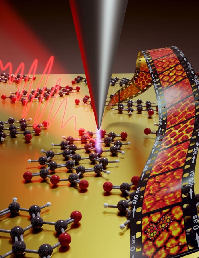
Tracking electron motion in molecules is the key to understanding and controlling chemical transformations. However, the goal of watching electron motion in real space and -time remains unrealized due to the limited resolution (in real space and -time) of existing techniques.
Contemporary techniques in attosecond science can generate and trace the consequences of this motion in real-time, but not in real space. On the other hand, scanning tunneling microscopy can locally probe the valence electron density in molecules but cannot provide dynamical information at this ultrafast timescale.
In a new study, Manish Garg and his colleagues from the Max Planck Institute for Solid State Research have combined scanning tunneling microscopy with ultrashort laser pulses and proposed a space-time quantum microscope. This space-time quantum microscope offers the desired space-time resolution to visualize electron motion directly.
The study could open new ways to understand chemical transformations driven by electron transfer. Plus, it could provide new insights, especially in charge transfer processes, which play a decisive role in many biophysical reactions and solar cells and transistors.
Manish Garg, head of a research group at the Max Planck Institute for Solid State Research, said, “We have been working for several years to combine these two techniques in such a way that each can play to its strengths without bringing up its weaknesses.”
“To do this, we had to couple tried-and-tested scanning tunneling microscopy with state-of-the-art laser technology. With a scanning tunneling microscope, a wafer-thin, atomically thin tip moves just above a conductive surface.”
The quantum physical tunnel effect allows electrons to flow between the surface and the microscope’s tip, that too without any direct contact.
In the new microscopy technique, laser pulses regulate the tunnel current through targeted excitation of the electrons in the material.
Alberto Martin-Jimenez, who played a crucial role in the experiments, said, “This has to happen extremely quickly; otherwise, thermal effects will come into play and make the measurements impossible.”
© Manish Garg / MPI for Solid State Research
This newly developed instrument allows the team to see the electron motion in molecules directly. They observed that the ultra-short laser pulses prompt electrons in the molecule to jump between the different orbitals. This was noticeable in the tunnel current.
Highlights of the new technology included firing two pulses at a precise time interval, one after the other in quick succession with a slight delay to probe and scan it in the process. Repeating the process multiple times at time interval gives several images, showing the behavior of the electrons in this molecule with atomic precision. The fast laser pulses, therefore, provide information about the electron dynamics.
Garg said, “This enabled us for the first time to directly map the dynamics of the electrons in molecules as they jump from one orbital to another. This basic technology provides completely new possibilities for directly observing quantum mechanical processes such as charge transfer in individual molecules and thus for better understanding them.”
The study also demonstrated concurrent real-space and real-time imaging of coherences involving the valence orbitals of molecules and complete control over the population of the involved orbitals.
© Manish Garg / MPI for Solid State Research
Study quotes, “This approach opens the way to the unambiguous observation and manipulation of electron dynamics in complex molecular systems.”
“The capability to image electronic coherences involving valence electronic states in molecules both at their natural length and natural timescales open completely new avenues to understand chemical transformations driven by electron transfer; for example, in photosynthetic molecules and other light-harvesting molecules, and to the unambiguous observation of electron dynamics in complex molecular systems, two-dimensional materials, and superconductors.”
Journal Reference:
- Garg, M., Martin-Jimenez, A., Pisarra, M. et al. Real-space subfemtosecond imaging of quantum electronic coherences in molecules. Nature Photonics (2021). DOI: 10.1038/s41566-021-00929-1
