The greater part of the body’s calcium is put away as a mineral in bones, a precisely controlled measure of this synthetic component is transported into the cell, in its ionic shape, where it assumes an imperative part in administering cell capacities. Cells manage calcium take-up through unique pores, or channels, that open and close as required.
TRPV6 is a protein divert that is situated in the films of epithelial cells, which line the dividers of the digestive tract and adds to the take-up of dietary calcium. Deviations in TRPV6 channels may add to the advancement of malignancy by upsetting the control of cell multiplication and cell demise.
Now, scientists at the Columbia University Medical Center (CUMC) have captured the first detailed snapshots of the structure of a membrane pore that enables epithelial cells to absorb calcium. Scientists believe that these images could optimize the development of drugs to correct abnormalities in calcium uptake, which have been linked to cancers of the breast, endometrium, prostate, and colon.
Scientists used advanced cryo-electron microscopy to image TRPV6. The Cryo-electron microscopy is an imaging technique that combines thousands of individual images of frozen molecules into finely detailed three-dimensional representations.
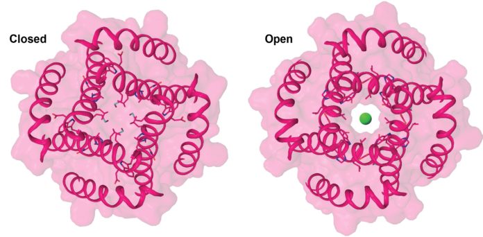
By contrasting the channel structures, in both the open and shut states, the scientists could verify that the center bit of the channel—four firmly adjusted helical protein fragments—complete an inconspicuous turn, enabling TRPV6 to open.
Study leader Alexander I. Sobolevsky said, “We discovered that the calcium channel opens in response to changes in the middle portion of each core helix, causing the protein segments to bend and rotate outward to create an opening just wide enough to allow a calcium ion to pass through.”
“If one were able to look straight down the channel, it would look like the opening of the iris of an eye.”
“Our findings will help us better understand how changes in TRPV6 channels contribute to human diseases such as cancer and provide a template for the design of drugs that correct these abnormalities.”
The channel can switch between open and closed states extremely quickly, as needed to supply the cell with calcium.
