
In a recent study, scientists have suggested that if a shoulder muscle appears unusually bright on ultrasound, it may be a warning sign of diabetes. The study which is being presented next week at the annual meeting of the Radiological Society of North America (RSNA), could allow for earlier interventions.
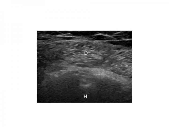
Ultrasound is normally used to analyze the source of pain in the shoulder. Over 10 years back, musculoskeletal radiologist Steven B. Soliman, D.O., from Henry Ford Hospital in Detroit, started seeing a pattern when pictures of the deltoid muscle, the biggest muscle of the shoulder, seemed bright on ultrasound.
The perceptions provoked Dr. Soliman and associates at Henry Ford to lead an investigation to check whether the brightness or echogenicity, of the shoulder muscle, could be prescient of diabetes.
CREDIT
Radiological Society of North America
The outcomes uncovered that by utilizing the echogenicity of the muscle, radiologists could foresee type 2 diabetes, the most well-known sort of diabetes, in very nearly nine out of 10 patients. Brightness on ultrasound likewise was an exact indicator of pre-diabetes, a state of anomalous high glucose that for the most part advances to diabetes without changes in way of life.
For the study, scientists compiled 137 shoulder ultrasounds from patients with type 2 diabetes, including 13 with pre-diabetes. The researchers also obtained 49 ultrasounds from obese patients without diabetes.

CREDIT
Radiological Society of North America
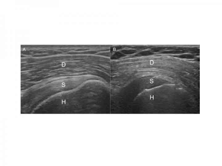
The researchers showed the ultrasounds to two musculoskeletal radiologists who were unaware of whether the images came from patients with or without diabetes. The radiologists were asked to classify the patients, based on the brightness of their shoulder muscle, into one of three categories: normal, suspected diabetes and definite diabetes. A third musculoskeletal radiologist acted as an arbitrator in the cases where the other two radiologists disagreed.
The results showed that a consensus diagnosis of “definite diabetes” by the radiologists was a powerful predictor of diabetic status. Using the shoulder ultrasounds, the radiologists correctly predicted diabetes in 70 of 79 patients or 89 percent.
Dr. Soliman said, “We weren’t surprised that we had positive results because the shoulder muscle on patients with diabetes looked so bright on ultrasound, but we were surprised at the level of accuracy.”
CREDIT
Radiological Society of North America
“A hyperechoic, or unusually bright-looking, deltoid muscle was also a strong predictor of pre-diabetes. The musculoskeletal radiologists assigned all 13 pre-diabetic ultrasounds to either the “suspected diabetes” or “definite diabetes” categories.”
“A lot of the patients weren’t even aware that they were diabetic or pre-diabetic. This lack of awareness is a major problem in the U.S.”
Dr. Soliman stated, “The reasons for the brighter-appearing shoulder muscle on ultrasound among patients with diabetes is not completely understood. But it is due to low levels of glycogen in the muscle, a key source of energy for the body that is stored primarily in the liver and muscles.”
An investigation of muscle biopsies in patients with diabetes discovered that muscle glycogen levels are diminished up to 65 percent. Earlier research has additionally demonstrated that the muscles of competitors seem more brilliant on ultrasound after exercise when their glycogen stores are drained.
Dr. Soliman said, “It could be that this appears in people with diabetes and pre-diabetes is related to the known problems with glycogen synthesis in their muscles because of their insulin abnormalities.”
If they see a bright shoulder muscle on ultrasound, radiologists at Henry Ford now put notes in their reports indicating that this observation has been linked to diabetes.
The researchers plan to continue studying the connection between shoulder muscle echogenicity and diabetes with an eye toward quantifying the phenomenon and seeing if it is reversible.