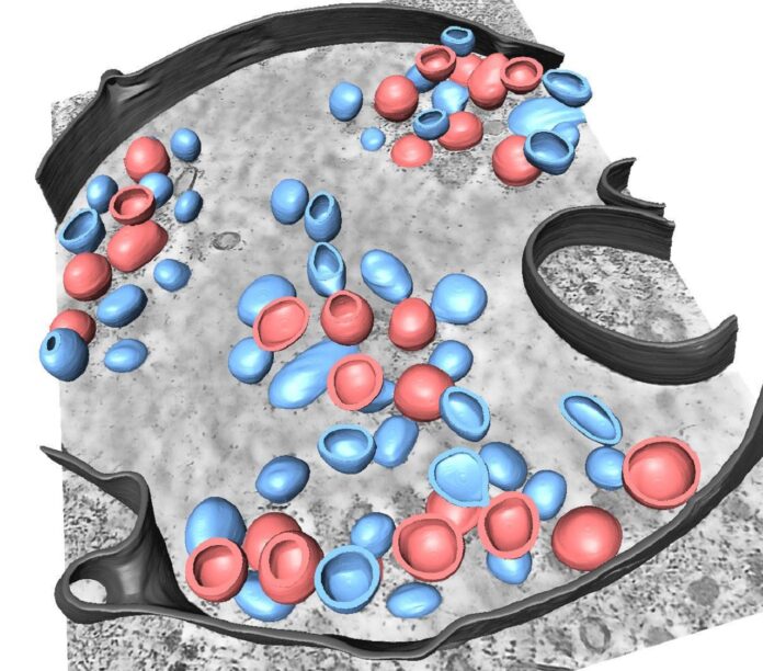
Attracting the virus into a trap is a concept that refers to the process of controlling the spread of a virus by attracting it to a specific area or environment where it can be easily contained and neutralized. This can be achieved through various methods, such as using bait to attract the virus to a designated area or creating a controlled environment that makes it difficult to escape.
Researchers from Heidelberg University and Heidelberg University Hospital used electron tomography and computer simulations to investigate the final stages of viral penetration for influenza A and Ebola viruses. They discovered that the immune system fights off influenza A using a small protein. For Ebola viruses, a specific protein structure must be disassembled for an infection to occur.
Fusion pores, through which the viral genome is released into the host cell, play a central role in these processes. Preventing the formation of fusion pores could block the virus, leading to new approaches to prevent infections.
Ebola viruses (EBOVs) cause severe viral hemorrhagic fever outbreaks in humans. They enter host cells through macropinocytosis and undergo cytosolic entry in late endosomal compartments. VP40 drives the budding of EBOVs and is critical for incorporating the viral nucleocapsid.
The nucleocapsid is composed of NP, VP24, and VP35. Fusion of the viral and endosomal membrane relies on interactions with the EBOV fusion glycoprotein (GP). Matrix disassembly during viral entry can trigger a cascade of events required for viral uncoating and efficient virus entry. VP40 and its interactions with lipids in the viral envelope are sensitive to low pH, which leads to the disassembly of the matrix layer and allows for fusion and genome release.
The human immune system tries to prevent the fusion pore formation during a viral infection. The infected cells send a signal to uninfected cells using interferon molecules. The uninfected cells then produce a small protein called IFITM3, which helps fight the virus.
Dr. Petr Chlanda, whose working group belongs to the BioQuant Center of Heidelberg University and the Center for Integrative Infectious Disease Research of Heidelberg University Hospital, said, “This specialized protein can effectively prevent viruses such as influenza A, SARS-CoV-2, and Ebola from penetrating, but the underlying mechanisms were unknown.”
The researchers could now demonstrate that IFITM3 selectively sorts the lipids in the membrane locally for influenza A viruses. This prevents the fusion pores from forming. “The viruses are captured in a lipid trap. Our research indicates that they are ultimately destroyed,” explains Dr Chlanda.
Dr. Chlanda and his team used equipment from the Cryo-Electron Microscopy Network at Ruperto Carola to analyze the structural details of viruses. The researchers investigated the penetration and fusion of the Ebola virus. They discovered that the VP40 matrix protein layer disassembles at a low pH, enabling the virus to form fusion pores with the host cell membrane.
The research suggests that a blockade of this disassembly could prevent fusion pore formation and trap the Ebola virus. The studies were part of the Collaborative Research Centre “Integrative Analysis of Pathogen Replication and Spread.” They were published in “Cell Host & Microbe” and the “EMBO Journal.”
In this study, the researchers used cryo-electron microscopy, tomography, and computational simulations to study the VP40 matrix layer of the Ebola virus and its interactions with the virus envelope and endosomal membrane. They prepared the samples by infecting VeroE6 cells with the Ebola virus and then fixed and embedded them in a low-temperature resin for imaging.
The data obtained were processed using software packages such as IMOD, Relion, and ChimeraX. The simulations were carried out using different computational modeling software, including GROMACS and CHARMM. The study was part of the Collaborative Research Centre “Integrative Analysis of Pathogen Replication and Spread” (CRC 1129), funded by the German Research Foundation.
The study discovered that the VP40 matrix layer of the Ebola virus disassembles at low pH in the endosomal membrane, which is crucial for the virus to fuse with the host cell membrane. Cryo-electron microscopy, tomography, and computational simulations were used to study the interactions between the virus envelope, the VP40 matrix layer, and the endosomal membrane.
The disassembly of the VP40 matrix layer weakens its electrostatic interactions with the membrane, reducing the energy barrier of pore formation required for the virus to release its genome into the host cell. The study proposes blocking the disassembly of the VP40 matrix layer as a potential antiviral therapy for the Ebola virus.
In conclusion, the Ebola virus VP40 matrix layer is an essential component of the virus’s structure that undergoes endosomal disassembly to facilitate membrane fusion.
Understanding the mechanisms by which the virus interacts with and enters host cells is crucial for developing effective treatments and vaccines. Further research in this area may lead to new strategies for preventing and controlling outbreaks of Ebola and other related viruses.
Attracting the virus into a trap involves capturing and controlling viruses to prevent their spread. Examples include using mosquito traps to control mosquito-borne illnesses and creating controlled environments such as quarantine centers and negative-pressure rooms for COVID-19.
Personal protective equipment and advanced technology such as AI and machine learning are also being used to prevent virus spread. This concept has great potential for protecting public health.
Journal Reference:
- Sophie L Winter, Gonen Golani,Fabio Lolicato, metal. The Ebola virus VP40 matrix layer undergoes endosomal disassembly essential for membrane fusion. EMBO. DOI: 10.15252/emj.2023113578
