Until recently, it was thought that quiet synapses were only present in the early stages of brain development when they assisted the brain in assimilating new information. However, the most recent MIT study found that around 30% of all synapses in the cortex of adult mice’s brains are silent. Scientists also found that these silent synapses remain inactive until they’re recruited to help form new memories.
These silent synapses may provide insight into how the adult brain can continuously create new memories and acquire new information without needing to change its old conventional synapses.
Dimitra Vardalaki, an MIT graduate student and the lead author of the new study, said, “These silent synapses are looking for new connections, and when important new information is presented, connections between the relevant neurons are strengthened. This lets the brain create new memories without overwriting the important memories stored in mature synapses, which are harder to change.”
In this study, scientists did not set out expressly to look for silent synapses. Instead, they were investigating a fascinating result from a prior experiment in Harnett’s lab (Mark Harnett, an associate professor of brain and cognitive sciences). In that study, the authors showed how dendrites, which resemble antennae and sprout from neurons, can process synaptic input differently depending on where they are located within a single neuron.
To determine if this could help explain the variations in their behavior, the scientists attempted to quantify the neurotransmitter receptors in various dendritic branches as part of that study. They achieved this using a method known as IMAP (epitope-preserving Magnified Analysis of the Proteome), which Chung created. This method allows for the physical expansion of tissue samples, followed by labeling specific proteins to provide images with extremely high resolution.
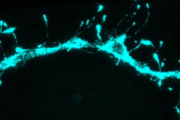
Harnett said, “While we were doing imaging, we y made a surprising discovery. The first thing we saw, which was super bizarre and we didn’t expect, was filopodia everywhere.”
Filopodia are thin membrane protrusions that extend from dendrites. Scientists have seen them earlier, but what they do remains unclear because they are so tiny that they are difficult to see using traditional imaging techniques.
Following this discovery, the MIT team used the IMAP technology to search for filopodia in additional adult brain regions. To their amazement, they discovered filopodia at a level 10 times higher than previously observed in the mouse visual cortex and other regions of the brain. Additionally, they found that filopodia lacked AMPA receptors but did have NMDA receptors, which are neurotransmitter receptors.
A typical active synapse has both of these receptors, binding to the neurotransmitter glutamate. NMDA receptors normally require cooperation with AMPA receptors to pass signals because magnesium ions block NMDA receptors at the normal resting potential of neurons. Thus, when AMPA receptors are absent, synapses that have only NMDA receptors cannot pass along an electric current and are referred to as “silent.”
The scientists adapted the experimental method known as patch clamping to look into the possibility that these filopodia are silent synapses. By simulating the release of the neurotransmitter glutamate from a nearby neuron, they could stimulate specific filopodia while simultaneously monitoring the electrical activity produced there.
Scientists used the technique to find that glutamate would not generate any electrical signal in the filopodium receiving the input unless the NMDA receptors were experimentally unblocked.
Scientists noted, “This strongly supports the theory that filopodia represent silent synapses within the brain.”
By combining glutamate release with an electrical current coming from the neuron’s body, it is possible to unsilence these silent synapses, showed scientists. This combined stimulation leads to the accumulation of AMPA receptors in the silent synapse, allowing it to form a strong connection with the nearby axon releasing glutamate.
The interesting fact is that converting these silent synapses into active ones was much easier than altering mature synapses.
Harnett said, “If you start with an already functional synapse, that plasticity protocol doesn’t work. The synapses in the adult brain have a much higher threshold, presumably because you want those memories to be pretty resilient. You don’t want them constantly being overwritten. Filopodia, on the other hand, can be captured to form new memories.”
“This paper is, as far as I know, the first real evidence of how it works in a mammalian brain. Filopodia allow a memory system to be both flexible and robust. You need the flexibility to acquire new information but also stability to retain the important information.”
The search for these quiet synapses in human brain tissue is currently underway. Additionally, they want to investigate how aging and neurodegenerative diseases, as well as other variables, may influence the number or functionality of these synapses.
Harnett said, “It’s entirely possible that by changing the amount of flexibility you’ve got in a memory system, it could become much harder to change your behaviors and habits or incorporate new information. You could also imagine finding some of the molecular players that are involved in filopodia and trying to manipulate some of those things to try to restore flexible memory as we age.”
Journal Reference:
- Vardalaki, D., Chung, K. & Harnett, M.T. Filopodia are a structural substrate for silent synapses in adult neocortex. Nature, 2022 DOI: 10.1038/s41586-022-05483-6
